Resources for those not getting adequate treatment

Hi, I’m 26F and have been dealing with intermediate uveitis since May 2023. I’ve had a trickle down of neurological symptoms since March 2023, new/worsening symptoms over time are severely affecting me and my life. I was on a low dose of prednisone up until October 2023, which helped subside the eye inflammation but did not clear it and I experienced new eye symptoms while I was on it. I am still having disruptive floaters/snow vision, fireworks/glittering, blur/haze, sometimes sharp pain and double vision.

Lumbar Puncture in July 2023 with lymphocytic pleocytosis, elevated CSF igG index, CSF Oligoclonal bands. Repeat LP in October 2023 revealed 6x higher inflammation, negative for infectious viral/bacterial/fungal causes. MRI with small non-specific non-enhancing bilateral subcortical hyperintensities in frontal lobes as of August 2023. Clear thoracic/cervical spine MRI as of August 2023. No blood inflammation or abnormalities other than elevated beta2glycoprotein antibodies. No abnormalities on chest/abdomen/pelvis CT. No previous medical conditions, events, surgeries or hospitalizations.

I have not had any testing on my eyes other than photography.

Doctors are currently unwilling to provide further treatment for the inflammatory immune response that I am having because they don’t know the ‘exact cause’ and because my findings/symptoms do not fit clearly inside of a diagnostic box. I am just getting worse and my symptoms are not improving at all. I’m not able to live a normal life and don’t know what to do/where to go from here – I have sought multiple opinions from various specialists. I am on state Medicaid insurance, so I cannot afford to see anyone outside of my state.

Are there any resources available for this type of situation or universities/clinics that would be willing to evaluate me as a patient outside of my state (VA) that would be more knowledgeable/better equipped to deal with a complex case?

Answer: Hi, so sorry to hear your story of obstacles to care.  I think this is more of a clinical question and needs to some clarification on your history so I would ask that you provide your email so we can communicate.  Please use the conus link on uveitis.org and email the support group link.  Thank you.  Frances Foster NP

ESSENTIAL Medical Visits requiring an Office Appointment

COVID19 Patient Information Update for MERSI Eye Center and OIUF

As you may have heard, Governor Baker has issued “A Stay in Place Order” for the state of Massachusetts for all non-essential businesses beginning at noon on March 24, 2020.   MERSI is considered essential, but we will need to restrict visits to those defined as essential.

ESSENTIAL Medical Visits requiring an Office Appointment:

  • Any patient actively being treated with immunosuppressive therapy (oral, infusion, or injection)
  • Any patient with acute or new symptoms/problems
  • Any follow up patient who had acute or new symptoms/problems and was placed on active treatment needing reassessment
  • Patients who are experiencing flashers, floaters, eye pain, or light sensitivity
  • Retina patients who are receiving injections to control their eye disease progression
  • Glaucoma patients actively being treated for elevated pressure issues or in late-stage disease
  • If you  feel the situation is urgent, call to schedule an appointment or arrange for a phone call with one of our clinical staff to determine the nature of the urgency for the visit
  • All routine, annual, screening appointments will be rescheduled unless you have new symptoms (which can be discussed with one of our clinical staff to determine urgency)

****We ask if you are sick or have returned from travel or been exposed to someone from who had traveled or is high-risk for contracting the Covid 19 virus to reschedule your appointments.***

Recommendations for those of you on immunomodulatory therapy:

We recommend you always  be vigilant about practicing universal precautions to prevent getting ill (see below).  The flu is a concern each year, which is why we recommend and encourage you to get the flu shot and now, we have a new viral illness in addition to the flu that we all are at risk for contracting.

Coronavirus disease (COVID-19) is an infectious disease caused by a new virus that had not been previously identified in humans.

The virus causes respiratory illness (like the flu) with symptoms such as a cough, fever and in more severe cases, pneumonia.

HOW IT SPREADS:

The new coronavirus like the flu spreads primarily through contact with an infected person when they cough or sneeze, or through droplets of saliva or discharge from the nose.

Practices to Protect Yourself:

  1. We encourage universal precautions, which one should practice always to avoid the flu, cold, or other illnesses in addition to the Coronavirus.
    1. 20-30 second handwashing, use hand sanitizer if no soap after touching surfaces and always before eating
    2. disinfect surfaces you will need to touch including your work space
    3. avoid touching door knobs or handles, elevator buttons, shopping carts, etc with hands. Use instead for example a wipe, paper towel, shirt sleeve, glove, or on elevator button your knuckle instead of finger tip  
    4. do not eat food with hands  
    5. avoid touching your face, mouth, and nose with hands
  • Air Travel or public spaces Postpone to avoid any risks until the virus spread is under control. 
  • The CDC provides great guidelines on their website. Please visit www.cdc.gov

Of note, If you develop any illness, infection, or fever of 99 degrees F or > or if you have been exposed to a high risk person for Covid 19 or Flu; please notify your doctor immediately and hold your immunosuppressive therapy medication immediately.  Specifically, if you have a fever of 101° Fahrenheit (38.3° Celsius), with or without chills, call your doctor immediately.   If you cannot reach your doctor, go to an emergency room.

In general, when you are healthy your risk for contracting an illness lessens so we recommend 8 hours of sleep, good hydration (8-10 glasses of noncaffeinated fluid a day), a multivitamin daily, and exercise if possible 3 times a week.   We also recommend smoking cessation if applicable to you.

Mersi Providers and Nursing Staff


Recent Research Relating to Cicatricial Pemphigoid and the Use of Intravenous Immunoglobulin (IVIG)

C. Stephen Foster, MD, FACS, FACR

Dr. Foster et al. recently published a chart review of all Ocular Cicatricial Pemphigoid (OCP) patients seen at the Massachusetts Eye Research and Surgery Institution (MERSI) between 2005 and 2015 to look at the management of OCP with Intravenous Immunoglobulin (IVIG) as the only therapy. IVIG infusion was administered in the usual manner with the dose being two grams per KG of weight and the total dose divided for infusion over three consecutive days every month.

Five hundred and twelve (512) patients were identified with OCP at MERSI, and only17 patients or 34 eyes were treated with IVIG as the only therapy.  Seven were female, and ten were male with the average age at diagnosis as 60.7 years old.  The follow-up time ranged from 12 to 140 months. Twenty-six eyes (76.5%) achieved remission. Nine remission eyes received cataract surgeries, and 2 of them had a relapse (22.2%). The other 17 eyes did not undergo ocular surgery and remained in remission.

The findings revealed that IVIG as only therapy showed high efficacy in stage one on Foster staging scale of OCP (7/7, 100%).  Ocular surgery can be associated with OCP relapse.  The conclusion from the chart review study was IVIG monotherapy is an effective and safe therapy in patients with stubborn OCP.  However, ocular surgery can be associated with OCP relapse even when a patient was in remission.  Rituxan and IVIG combination are still top of the line treatment for OCP.

#ocularcicatricialpemphigoid #ocp #intravenousimmunoglobulin #IVIG

Cost of Care of Patients with Uveitis

C. Stephen Foster MD

Increasingly restrictive “gate keeping” policies of health maintenance organizations, insurance companies, and other medical insurance plans have created increasing pressure on ophthalmologists to be parsimonious in their use of medical services in both the diagnostic and therapeutic care of patients with a variety of medical disorders, including those with uveitis. These pressures are particularly prominent in the physician’s care of patients with chronic disorders, and ophthalmologists caring for patients with uveitis are increasingly experiencing this restrictive pressure. We wondered what the cost of diagnostic and therapeutic care of a patient with uveitis might be, given what we, as a uveitis referral center, see as appropriate yet fiscally prudent care. The cost of care obviously varies greatly, depending on the underlying cause and on the severity of the patient’s uveitis and associated complications. As a first step in estimating the total annual direct cost in the care of patients with uveitis we restricted our analysis to patients with HLA-B27 associated uveitis. We also restricted our analysis to the direct medical cost of caring for such patients, recognizing that direct non-medical costs, indirect morbidity costs, and other intangible economic loss costs, disability payments, absences from work, etc. are real but difficult to measure costs of the total cost of the patient’s illness. Direct medical costs are transactions and expenditures for medical products and services, including diagnostic studies, physician fees, hospitalization costs, surgical costs, rehabilitation and subsequent long-term care costs.

A cohort of 105 patients with HLA-B27 associated uveitis were studied in 2002. The diagnosis in each instance was established on our Service, and a minimum follow-up of two years existed for each patient. The medical records were reviewed for the diagnostic studies and costs of each performed, the physician and hospital fees associated with visits and/or surgery, in the cost of medical therapy. The average direct annual cost per patient per year was calculated. A stepladder approach to therapy was employed in an effort to eliminate recurrences of uveitis. The first step on the stepladder was the use of steroids, through any route required to achieve the goal of quieting the uveitis. Oral non-steroidal anti-inflammatory agents were added, if recurrence typically continued despite the use of steroids. Immunosuppressive chemotherapy was employed if patients continued to have recurrence of inflammation despite the use oral non-steroidals. Ten patients eventually required the use long-term oral immunosuppressive agents, and 30 patients were on chronic oral non-steroidal anti-inflammatory drugs. The average annual cost of care of the patients with HLA-B27 associated uveitis was $4,108.60 (range $433 to $9,683.18). These results reflect an average cost of caring for a cohort of patients with recurrent HLA-B27 associated uveitis of varying severity. The results may serve as an indicator, to health maintenance organizations and other pooled-risk insurers, of the cost of prudent care of patients with this form of uveitis. We would emphasize that we were very cautious and parsimonious in our use of laboratory tests and frequency of return visits, striving for the greatest degree of economy, while at the same time striving for the best possible outcomes (for outcomes analysis studies performed on these and other patients with uveitis, please refer to the Bibliography section of this Web Site). Clearly, patients with recurrent or chronic uveitis require significant expenditure of the health care dollar. It is, however, money well spent, since the preservation of sight from modern care of such patients profoundly reduces the prevalence of blindness secondary to uveitis, and hence reduces the economic burden on our society in total.

Did you know the Connection between Arthritis and Ocular Disease?

Connection Between Arthritis and Ocular Disease
C. Stephen Foster, M.D.

The eye is made up primarily of collagen, as are ligaments, tendons, and tissue within joint spaces. It is, perhaps, primarily because of this similarity in composition that the eye is often affected by many of the same diseases which affect joints. Some of these disorders include Juvenile Rheumatoid Arthritis, Adult Rheumatoid Arthritis, Systemic Lupus Erythematosus, Relapsing Polycondritis, Behcet’s Disease, Granulomatosis with Polyangiitis (formerly called Wegener’s), Polyarteritis Nodosa, and Scleroderma or systemic sclerosis. Additionally, the type of vasculature that is present in the eye has special characteristics that produce an extraordinarily sensitive “barometer” or “sentinel canary” in the eye for potentially lethal vasculitis that can be associated with the aforementioned collagen vascular diseases. Specifically, we know from considerable experience that, despite the fact that a patient’s rheumatoid arthritis may be “burned out” as far as active inflammation of the joints in concerned, nonetheless, the patient may well have subclinical rheumatoid vasculitis affecting various internal organ systems. The eye is a very potent indicator of such subclinical potentially lethal vasculitis, and if the eye becomes involved with retinal vasculitis, uveitis, scleritis, or peripheral ulcerative keratitis in such a patient, we take that as a very strong signal that the patient must be evaluated extremely carefully for potentially underlying vasculitis affecting viscera and we also take such a potentially blinding ocular lesion very seriously from the standpoint of the need for aggressive systemic immunomodulatory therapy in order to prevent permanent damage to the eye from such lesions.

For example, we have seen many instances in which patients with systemic lupus erythematosus appear, systemically, to be doing quite well (indeed, the patient’s Rheumatologist has told her that she is doing very well) despite the fact that new-onset uveitis, scleritis, or retinal vasculitis has developed in one eye. We have seen this story evolve to life-threatening central nervous system vasculitis and/or lupus renal disease when the onset of the ocular inflammation was not taken as an indication for increasing the vigor of systemic therapy. We have tried diligently, therefore, over the past 15 years to raise the consciousness, not only of ophthalmologists worldwide, but also of rheumatologists and other internists of the valuable indicator that the eye can be with respect to seriousness of associated arthritic/collagen vascular disease.

Pediatric Uveitis

Uveitis is the third leading cause of blindness in America, and 5% to 10% of the cases occur in children under the age of 16. But Uveitis in children blinds a larger percentage of those affected than in adults, since 40% of the cases occurring in children are posterior uveitis, compared to the 20% of posterior Uveitic cases in the adult Uveitis population.

There are, at any one time, approximately 115,000 cases of Pediatric Uveitis in the United States, with 2,250 new cases occurring each year. Spread across the entire U.S. population, therefore, and across all offices of Ophthalmic practitioners, the likelihood that any one individual practitioner will care for a patient with Pediatric Uveitis is relatively small, and the likelihood that any single individual will have significant experience in caring for large numbers of cases over a long period of time is vanishingly small. This accounts, we believe, at least in part for the sub-optimal care that many of our children with Uveitis appear to be receiving, even in these “modern” times. The stakes are incredibly high, for the child, for the parents who will be faced with (usually) many years of dealing with this health problem in their child, and for society at large because of the life-time of dependence which occurs in those who eventually reap substantial visual handicap as the result of sub-optimal treatment.

We believe that current epidemiologic data emphasize two critically important goals for all of us in Ophthalmology, acting together, in an effort to change the current prevalence of blindness caused by Pediatric Uveitis:

  1. Repeatedly emphasizing to parents, other medical colleagues, especially Pediatricians, and school personnel the critical importance of routine (annual) vision screening for all children.
  2. The critical importance of beating back the frontiers of general ignorance and mind sets, eliminating the all-too-common pronouncement by physicians to parents of a child with Pediatric Uveitis that:
  3. “He’ll (She’ll) out grow it.”
  4. “The drops will get him (her) through it.”
  5. “It’s just the eye; systemic therapy is not warranted.”

Statements (a) and (b) are true, but too often pull the doctor, and patient, and family into the seduction of nearly endless amounts of topical steroid therapy. It is generally true that the child will in fact “out grow” the Uveitis, i.e., that the Uveitis will no longer be a problem eventually. The pity is, however, that so often by the time the child “out grows it”, permanent structural damage to retina, optic nerve, or aqueous outflow pathways has already occurred, and the blinding consequences are now permanent. It is also true that for any individual episode of Uveitis, the steroid drops usually will get the patient through it. But the fact is that so many children with Pediatric Uveitis have recurrent episodes of Uveitis such that the cumulative damage caused by each episode of Uveitis and the steroid therapy for each episode eventually produces vision-robbing damage. And item (c) is simply the result of the common myopic viewpoint of Ophthalmologists: that it is just an eye problem, and therefore should simply be treated with eye medications. Nothing could be further from the truth! And unless and until large numbers of Ophthalmologists reframe this socially and epidemiologically important matter, the prevalence of blindness secondary to Pediatric Uveitis is not going to change.

The differential diagnosis of Pediatric Uveitis is relatively vast, and therefore the detective work required to properly pursue the underlying diagnosis is complex. The job can be slightly simplified by “playing the odds”, categorizing the case as carefully as possible into anterior non-granulomatous; anterior granulomatous; intermediate; posterior, with vasculitis; posterior, without vasculitis; and categorizing it into the general age groups of Infancy (0 to 2 years), Toddler-School Age (2 to 10 years), and Adolescence (10 to 20 years).

The most common etiologic groups in children segregated into these groups are shown in Tables 1-6

TABLE 1 (Anterior Non-Granulomatous)

Idiopathic

HLA-B27 associated

Juvenile Rheumatoid Arthritis

Ankylosing Spondylitis

Reactive Arthritis (formerly called Reiter’s syndrome) disease

Psoriasis

Inflammatory bowel disease

Nephritis

Systemic lupus erythematosus

Herpes Simplex virus

Lyme disease

Leukemia

Drug-induced

TABLE 2 (Anterior Granulomatous Uveitis)

Sarcoidosis

Inflammatory bowel disease

Syphilis

Herpes simplex virus

Tuberculosis

Bechet’s disease

Multiple Sclerosis

Fungal disease

Whipple’s disease

Leprosy

TABLE 3 (Intermediate Uveitis)

JRA

Pars Planitis

Multiple Sclerosis

Lyme disease

Sarcoidosis

TABLE 4 (Posterior Uveitis, without vasculitis)

Toxocariasis

Toxoplasmosis

Leukemia

Tuberculosis

Intraocular Foreign Body

Vogt-Koyanagi Harada Syndrome

TABLE 5 (POSTERIOR UVEITIS, with vasculitis)

Posterior Uveitis with vasculitis

Cytomegalovirus

HSV/VZV

Inflammatory bowel disease

Syphilis

Bechet’s disease

Systemic lupus erythematosus

Kowasaki’s disease

Sarcoidosis

Polyarteritis nodosa

Granulomatosis with Polyangiitis (formerly called Wegener’s)

TABLE 6 (most common causes of Uveitis in infants)

Herpes Simplex Virus

Toxocara

Congenital Loes

Retinoblastoma

TABLE 7 (most common causes of Uveitis in Toddlers/School Children)

Toxocariasis

Toxoplasmosis

Leukemia

Vogt-Koyanagi Harada Syndrome

Diffuse Unilateral Sclerosing Neuroretinitis

Juvenile Rheumatoid Arthritis

TABLE 8 (most common causes in Adolescents)

JRA

Pars Planitis

VKH

Toxoplasmosis

HLA-B27-associated sarcoidosis

Bechet’s disease

Intraocular Foreign Body

We believe that aggressive efforts should be made to uncover the underlying cause of Uveitis in any child. If the review of systems is negative and the patient has non-recurrent anterior granulomatous Uveitis, we would not do laboratory studies. However, if review of systems is positive, we would “follow the review of systems”.

For recurrent anterior non-granulomatous Uveitis we would obtain a complete blood count and urine analysis, ANA testing, HLA-B27 testing, and would “follow the review of systems”.

The diagnostic step ladder in a pediatric patient with anterior granulomatous Uveitis, recurrent or not, would include a CBC with urine analysis, and FTA-ABS testing, Lyme disease antibody and western block, PPD analysis, chest X-ray, ANA, and angiotensin converting enzyme determination. Chest CT, and Gallium scanning would be pursued if diagnosis of sarcoidosis was strongly suspected, and, of course as usual, we would “follow the review of systems positives”.

In a patient with intermediate Uveitis, all would deserve laboratory evaluation, including CBC, urine analysis, chest X-ray, FTA-ABS, ACE, PPD, Lyme, and ANA titers.

Any patients with posterior Uveitis would deserve an extensive vasculitis work-up, if vasculitis were present, and a search for “the usual suspects” with an eye to an infectious etiology, such as that producing a granuloma in the choroid in a patient with toxocariasis or Toxoplasmosis. An audiogram or lumbar puncture would be done if positive on the review of systems were found such as tinnitus and/or meningeal signs or symptoms. Finally, a diagnostic vitrectomy would be added to the step ladder in a patient with posterior Uveitis if all non-invasive studies were unrevealing and the case was difficult to treat successfully.

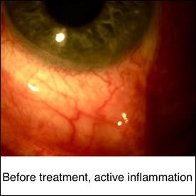
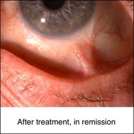
On the matter of treatment, here too we believe strongly in the step ladder approach, always beginning with steroids, in any route required to achieve the desired goal, i.e., abolition of all active inflammation. Topical steroids would be followed by an examination under anesthesia with regional steroid injection therapy in a patient with granulomatous or non-granulomatous anterior Uveitis. Systemic steroids would be employed in the event that this approach did not achieve the goal of abolition of all active cells. We are extremely reluctant to get involved with long term daily systemic steroid use in a youngster, because of the obvious growth-retarding properties of such therapy. But long term oral non-steroid anti-inflammatory drug therapy, managed by a Pediatrician, can be extremely successful, in our experience in approximately 70% of children with recurrent non-granulomatous anterior Uveitis. If this strategy is not successful, then consideration of once weekly, low-dose, Methotrexate or daily Cyclosporine or CellCept would be the next considerations.

In granulomatous disease topical steroids often are not sufficient, and systemic therapy, particularly with oral non-steroidal inflammatory drugs, may be utilized sooner rather than later.

With Intermediate Uveitis topical steroids are not effective in penetrating to the level of inflammatory focus. Regional steroid injections or systemic steroids are employed to treat that area, sometimes with adjunctive topical steroids for anterior chamber “spill over” reaction. Retinal Cyropexy can be effective in selected cases of recurrent Pars Planitis, as can therapeutic Pars Plana Vitrectomy. Systemic immunomodulatory therapy, as usual, represents the final step in the step ladder approach in the aggressiveness of care.

Patients with Posterior Uveitis of course do not respond to topical therapy and therefore require systemic steroids and/or immunomodulators right from the very beginning.

We hope that this will provide some help to those Ophthalmologists who have also concluded that the usual approach to Pediatric Uveitis, i.e., steroid drops, is not always sufficient, but who are hesitant to take the initiative to commit the patient to more aggressive treatment.